|
Input files
|
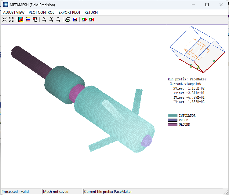
Pacemaker.MIN, Pacemaker.HIN, Pacemaker.SCR, Ground.OTL, Insulator01.OTL, Insulator02.OTL
Download Pacemaker3D.zip
|
|
Description
|
Determination of the electric field distribution and total current from a pacemaker inserted in heart muscle from a blood-filled chamber. A 5.0 V pulse of width 0.6 ms with rise and fall times of 0.1 ms is applied.
|
|
Results
|
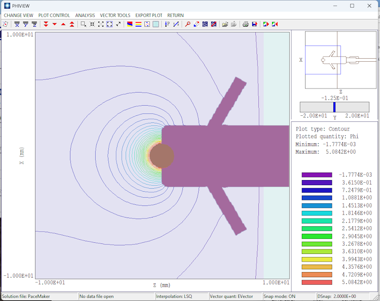
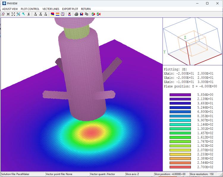
The reference Compilation of the Dielectric Properties of Body Tissues at RF and Microwave Frequencies (C. Gabriel, 1996) gives the following values for the electrical properties of the media: heart muscle (EpsiR = 2.0E5, Sigma = 0.1 S/m) and blood (EpsiR = 3000, Sigma = 0.7 S/m). Over a 0.1 ms risetime, displacement currents constitute less than 1% of current density, so flow is almost purely conductive. In this case, the voltage at all points in the solution space is proportional to the applied voltage, so a static electric-field solution is sufficient. The surface integral function of HiPhi shows that the probe releases a peak current of 3.057 mA into the muscle.
|
|
Comments
|
The geometry and parameters replicate those in the paper Finite element modeling of pacemaker electrode for time varying excitation (S. Kalra, M. Nabi, Proc. 32nd European Conference on Modeling and Simulation), represented by a set of five entries in the ComSol Application Gallery. The paper describes a time-dependent 3D calculation, although representation of temporal variations is not necessary. The effect of three-dimensional anchors on the device are small. A 2D model using the EStat code gives about the same released current, 2.932 mA.
|